Research
Spatial transcriptomic profiling uncovers the molecular effects of the neurotoxicant polychlorinated biphenyls (PCBs) in the brains of adult mice
Basu et al.
Molecular Psychiatry (Mol Psychiatry)
January, 2026
Abstract
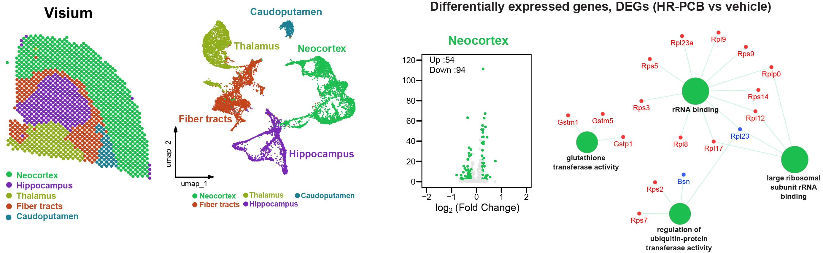
Polychlorinated biphenyls (PCBs) are highly stable synthetic organic compounds that are present in air, water, and soil. PCBs have been identified in post-mortem human brains, indicating a possible link between environmental factors and disease risk. Research has revealed an association between PCB exposure and cognitive decline. Therefore, it is crucial to evaluate how PCB mixtures relevant to humans affect brain function and cognition. To investigate the effects of PCBs on memory and transcriptomic profiles, we exposed male C57BL/6J mice orally to a synthetic PCB mixture daily. After seven weeks of exposure, the adult mice were assessed in a spatial object recognition task (SOR) to evaluate long-term spatial memory. Our findings showed that mice exposed to PCBs exhibited deficits in long-term spatial memory. To examine the molecular effects of PCB on the brain, we used a spatial transcriptomics technique to analyze gene expression changes in five brain regions: the hippocampus, neocortex, thalamus, caudal putamen, and fiber tracts. Our analysis of spatial gene expression revealed the molecular signatures influenced by PCB in these susceptible brain regions of mice. Network analysis suggests that these changes are associated with higher chlorinated PCBs present in the brain. Additionally, we show that PCB exposure disrupts the expression of tight junction proteins, which are crucial for maintaining the integrity of the blood-brain barrier (BBB). Thus, our results offer mechanistic insights into how PCB exposure affects brain function and cognition.
Differential Hes1 activation defines neural stem cell lineage commitment and niche maintenance in embryonic and adult mouse cortex
Riya et al.
Proceedings of the National Academy of Sciences (PNAS)
January, 2026
Abstract
Mode of Hes1 activation and its differential expression are crucial for the maintenance of neural stem cells/progenitor cells (NSCs/NPCs) in the embryonic cortex. This differential mode of Hes1 activation has been translated into a heterogeneous population of NSCs comprising Notch-independent Hes1-expressing (NIHes1) NSCs and Notch-dependent Hes1-expressing (NDHes1) radial glial cells (RGCs). Using single-cell transcriptomics and a Nestin-CreERT2;NIHes1fl/fl conditional knock-out mouse model, we have characterized the NIHes1 NSCs. Our analyses show that NIHes1 NSCs are the ancestral precursor NSCs that generate RGCs and intermediate progenitor cells during development. Loss of NIHes1 expression significantly alters the NSC niche, leading to increased gliogenesis and aberrant migration of projection neurons. NIHes1 NSCs are set aside at embryonic stages as adult neural stem cells and are maintained by NIHes1 expression even at adult stage. Our findings suggest that NIHes1 NSCs are functionally distinct Hes1-expressing NSCs, which are critical for establishing both embryonic and adult NSC niches, thereby contributing to the overall cortical development.
Single-cell resolution spatial transcriptomic signature of the retrosplenial cortex during memory consolidation
Bliese#,Basu# et al.
Molecular Psychiatry (Mol Psychiatry)
November, 2025
Abstract
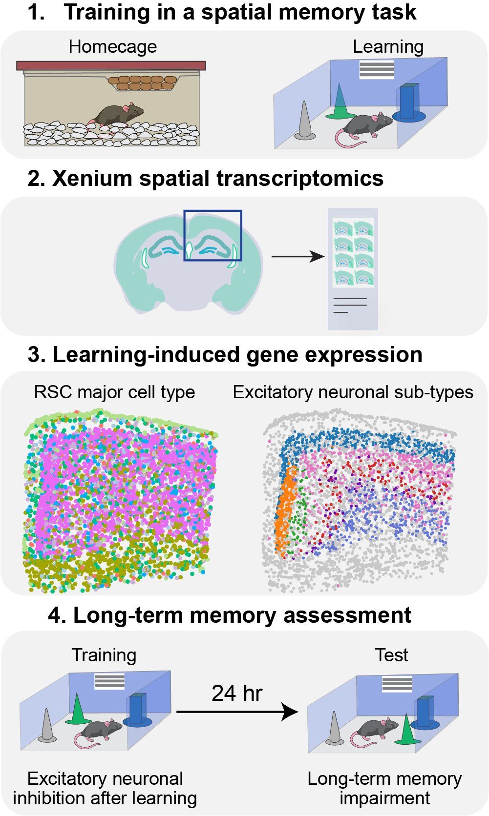
The retrosplenial cortex (RSC) is a critical brain region that is activated during spatial memory tasks and plays a crucial role in the consolidation of long-term memory. Various classes of RSC excitatory neurons across different laminar layers serve as the central hub for neuronal connections between the RSC and other brain regions, such as the hippocampus. Despite the established role of the RSC in spatial memory, the transcriptomic signature of the neuronal subtypes in the RSC during spatial memory consolidation remained elusive. Here, we used unbiased and targeted spatial transcriptomics to identify the RSC transcriptional signature after a spatial memory task. Genes related to transcription regulation, protein folding, and mitogen-activated protein kinase pathways were upregulated in the RSC during an early time window of memory consolidation. Furthermore, cell-type and excitatory neuronal layer-specific changes in gene expression were resolved using Xenium spatial transcriptomics. A deep learning computational tool uncovered cell-type-specific molecular activation patterns within the RSC after learning. Conversely, in a mouse model of Alzheimer’s disease and related dementia (ADRD) exhibiting tau hyperphosphorylation in the RSC, there was a reduction in predicted neuronal activation following learning. Notably, learning-induced Fos expression was decreased in excitatory neurons of the RSC in the ADRD mice. Finally, we observed that blocking RSC excitatory neurons during the early temporal window after learning using a chemogenetic approach impaired long-term spatial memory in adult mice. Our results reveal a molecular signature of the RSC after learning and emphasize the role of RSC excitatory neurons during spatial memory consolidation.
TLX3 regulates CGN progenitor proliferation during cerebellum development and its dysfunction can lead to autism
Parvathy et al.
iScience
Volume 27, Issue 12
December, 2024
Abstract
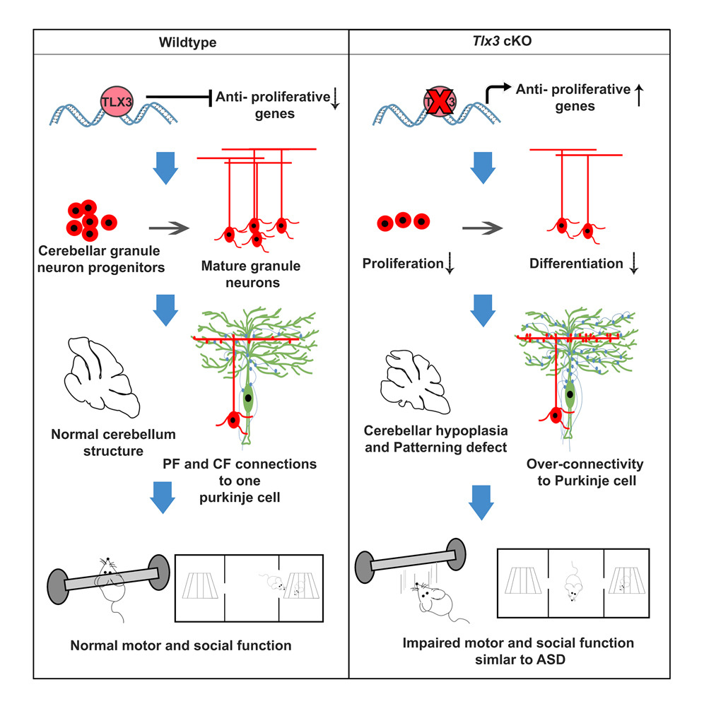
Tlx3, a master regulator of the fate specification of excitatory neurons, is primarily known to function in post-mitotic cells. Although we have previously identified TLX3 expression in the proliferating granule neuron progenitors (GNPs) of cerebellum, its primary role is unknown. Here, we demonstrate that the dysfunction of Tlx3 from the GNPs significantly reduced its proliferation through regulating anti-proliferative genes. Consequently, the altered generation of GNPs resulted in cerebellar hypoplasia, patterning defects, granule neuron-Purkinje ratio imbalance, and aberrant synaptic connections in the cerebellum. This altered cerebellar homeostasis manifested into a typical autism-like behavior in mice with motor, and social function disabilities. We also show the presence of TLX3 variants with uncharacterized mutations in human cases of autism spectrum disorder (ASD). Altogether, our study establishes Tlx3 as a critical gene involved in developing GNPs and that its deletion from the early developmental stage culminates in autism.
HES1 promoter activation dynamics reveal the plasticity,stemness and heterogeneity in neuroblastoma cancer stem cells
Riya#,Basu# et al.
Journal of Cell Science
Volume 135, Issue 22
November, 2022
Abstract
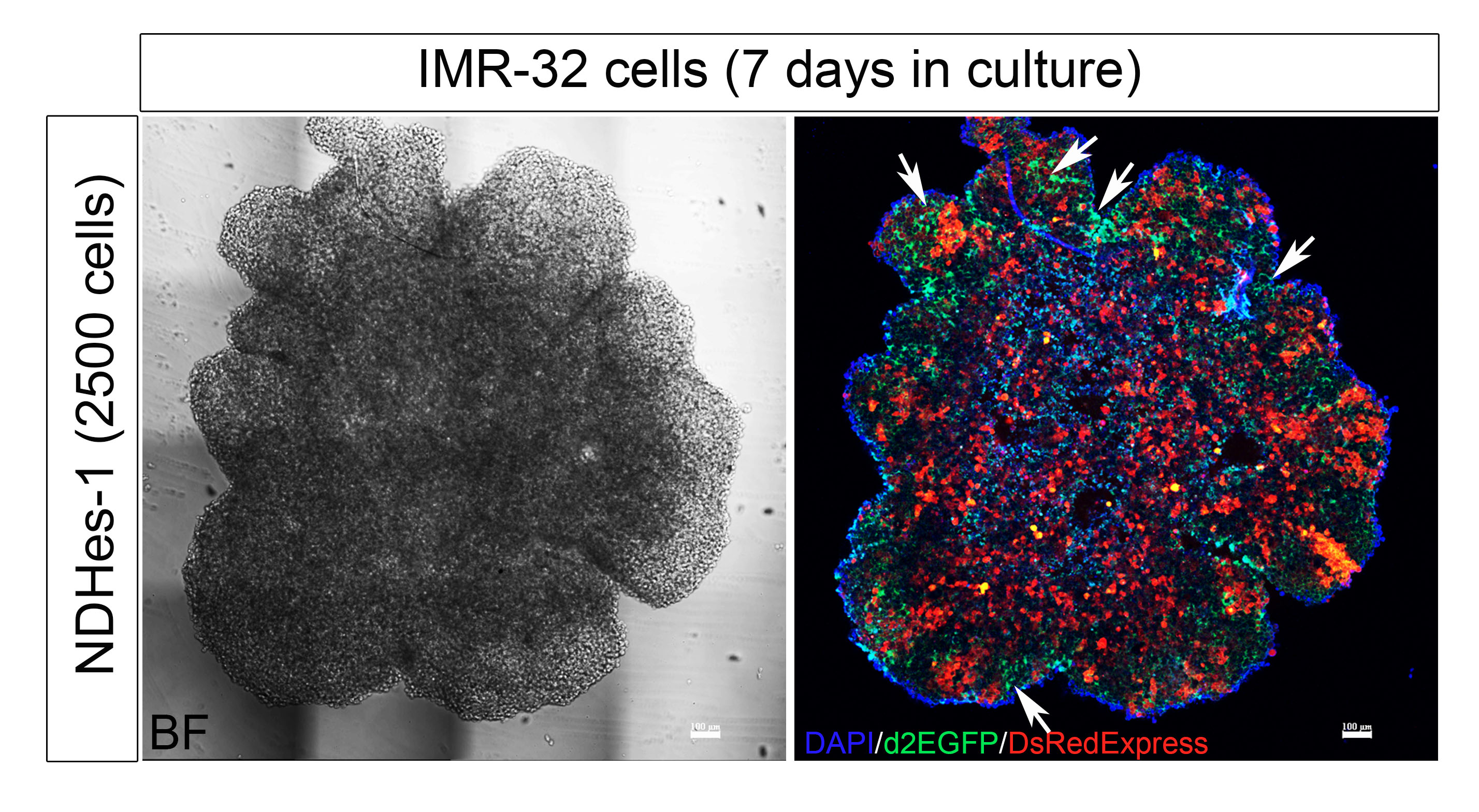
Notch signaling and its downstream gene target HES1 play a critical role in regulating and maintaining cancer stem cells (CSCs), similar to as they do during embryonic development. Here, we report a unique subclass of Notch-independent Hes-1 (NIHes-1)-expressing CSCs in neuroblastoma. These CSCs maintain sustained HES1 expression by activation of HES1 promoter region upstream of classical CBF-1 binding sites, thereby completely bypassing Notch receptor-mediated activation. These stem cells have self-renewal ability and potential to generate tumors. Interestingly, we observed that NIHes-1 CSCs could transition to Notch-dependent Hes-1-expressing (NDHes-1) CSCs where HES1 is expressed by Notch receptor-mediated promoter activation. We observed that NDHes-1-expressing CSCs also had the potential to transition to NIHes-1 CSCs and during this coordinated bidirectional transition, both CSCs gave rise to the majority of the bulk cancer cells, which had an inactive HES1 promoter (PIHes-1). A few of these PIHes-1 cells were capable of reverting into a CSC state. These findings explain the existence of a heterogenic mode of HES1 promoter activation within the IMR-32 neuroblastoma cell line and the potential to switch between them.
Pax6 modulates intra-retinal axon guidance and fasciculation of retinal ganglion cells during retinogenesis
Lalitha et al.
Scientific Reports
Volume 10, Issue 1
September, 2020
Abstract
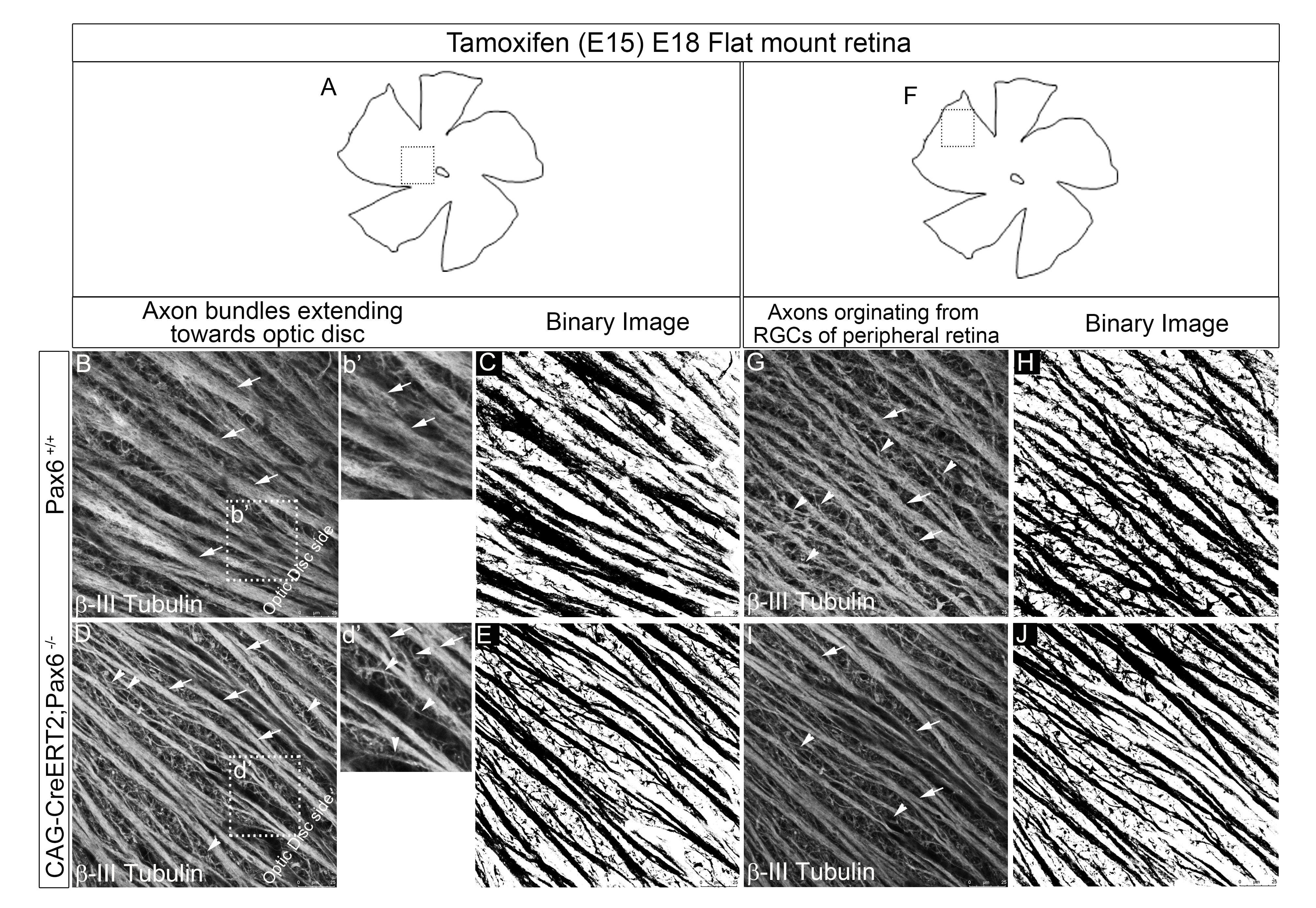
Intra-retinal axon guidance involves a coordinated expression of transcription factors, axon guidance genes, and secretory molecules within the retina. Pax6, the master regulator gene, has a spatio-temporal expression typically restricted till neurogenesis and fate-specification. However, our observation of persistent expression of Pax6 in mature RGCs led us to hypothesize that Pax6 could play a major role in axon guidance after fate specification. Here, we found significant alteration in intra-retinal axon guidance and fasciculation upon knocking out of Pax6 in E15.5 retina. Through unbiased transcriptome profiling between Pax6fl/fl and Pax6−/− retinas, we revealed the mechanistic insight of its role in axon guidance. Our results showed a significant increase in the expression of extracellular matrix molecules and decreased expression of retinal fate specification and neuron projection guidance molecules. Additionally, we found that EphB1 and Sema5B are directly regulated by Pax6 owing to the guidance defects and improper fasciculation of axons. We conclude that Pax6 expression post fate specification of RGCs is necessary for regulating the expression of axon guidance genes and most importantly for maintaining a conducive ECM through which the nascent axons get guided and fasciculate to reach the optic disc.